An M13 phage based colorometric sensor

Having previously demonstrated the ability to build collagen-like self-templating supramolecular structures out of M13 phage filaments, this year the Seung-Wuk Lee Lab has come out with a new paper demonstrating their ability to use those structures as a litmus to sensitively detect things like humidity, volatile organic compounds, or TNT explosives and report color strongly enough for an iPhone camera to distinguish:

Biomimetic virus-based colourimetric sensors
Many materials in nature change colours in response to stimuli, making them attractive for use as sensor platform. However, both natural materials and their synthetic analogues lack selectivity towards specific chemicals, and introducing such selectivity remains a challenge. Here we report the self-assembly of genetically engineered viruses (M13 phage) into target-specific, colourimetric biosensors. The sensors are composed of phage-bundle nanostructures and exhibit viewing-angle independent colour, similar to collagen structures in turkey skin. On exposure to various volatile organic chemicals, the structures rapidly swell and undergo distinct colour changes. Furthermore, sensors composed of phage displaying trinitrotoluene (TNT)-binding peptide motifs identified from a phage display selectively distinguish TNT down to 300 p.p.b. over similarly structured chemicals. Our tunable, colourimetric sensors can be useful for the detection of a variety of harmful toxicants and pathogens to protect human health and national security.

rsz_1ncomms4043-f1
“Bioinspired phage-based colourimetric sensors, termed Phage litmus, are composed of hierarchical bundles like the collagen fibres in turkey skins. Application of target molecules (chemical stimuli) causes colour shifts due to structural changes, such as bundle spacing (d1 and d2) and coherent scattering. Using a handheld device’s camera (iPhone) and home-built software (iColour Analyser), we can identify target molecules in a selective and sensitive manner.”

Ambiguous Phage Terms

Stephen T. Abedon

Department of Microbiology – The Ohio State University

phage.org – phage-therapy.org – biologyaspoetry.org


All fields employ specialized terms and at a minimum it is helpful for those individuals working in a field to both know and agree upon what those terms mean. As no doubt is also the case for most or all other fields, in phage biology there are a number terms that nonetheless possess ambiguous meanings. Here I provide both a list and brief discussion of my personal top-ten list of ambiguously defined or otherwise improper phage terms. Note that in many cases it generally is good practice to be aware of and then define ambiguous terms as you use them; this is so that your reader will understand what specific meaning you may be hoping to convey. Here then, in alphabetical order, is my list of top-ten ambiguous phage terms and why I’ve placed them on the list.

  1. Adsorption – This term is not so much ambiguous as potentially covering far too much ground. It can be used to describe the entire process of phage acquisition of a host bacterium, from diffusion through collision with a bacterium, attachment, virion conformational change, and even nucleic acid translocation. Alternatively, it can just mean attachment, though even that can be reversible attachment versus irreversible. In this case actually defining your intended meaning is not necessarily important, though keeping in mind the term’s ambiguous nature can’t hurt.
  2. Capsid – Though scientifically I “grew up” considering the entire phage particle sans the nucleic acid – and sans also the envelope, if present – as the capsid, in fact the capsid can be distinguished, in tailed phages, from the tail. The capsid thus surrounds and serves to contain and protect the nucleic acid and can contrast with other proteinaceous virion appendages which have other functions such as phage delivery into the adsorbed host cell.
  3. Carrier state – Different sub-fields use this term differently. Indeed, almost everybody uses this term with different meanings. If somebody says to you, “Carrier state”, you probably will assume that the intended meaning is whatever it is that you typically think the intended meaning should be. A little piece of advice: Don’t bet large amounts of money on that assumption.
  4. Lysis from without – Lysis from without is a term that almost makes me want to cry. There generally are four definitions used for the term, two of them both correct and distinct and two of them simply are wrong. If a phage particle, particularly when applied in high densities, lyses a target bacterium and does so well prior to the normal end of that phage’s latent period, then that’s lysis from without. If an endolysin is purified and then applied to a bacterium externally, resulting in lysis, then that also is lysis from without. By contrast, if you add large numbers of phages to a bacterium and the bacterium dies, that has almost no meaning except that phages can kill bacteria. As for the fourth usage, if you observe confluent clearing in the course of a spot test, then that’s a zone of inhibition rather than necessarily lysis from without, just like the zones of inhibition that antibiotics produce. Spot formation in fact says absolutely nothing about the lytic behavior of the phage applied other than that the phage in the numbers applied, or even the carrying fluid, can appreciably kill the target bacteria.
  5. Lysogenic phage – Bacteria are lysogenic. That is, if they contain a prophage then they have the potential to generate lysis in a second bacterial strain following the mixing of cultures. What people mean to say when they say lysogenic phage is temperate phage. Lysogenic phage is ambiguous in the sense that it is a misapplied term. Please, just don’t use it.
  6. Lytic phage – So, what is a lytic phage? A phage that lyses bacteria? What kind of information does that supply? That it isn’t a chronically released phage? Is that the intended meaning when “lytic” is used as a qualifier for “phage”? Sometimes, yes it is. Usually, though, the term lytic phage seems to be used to mean non-temperate. The logic of this meaning, however, is not necessarily well worked out since most temperate phages technically are also lytic phages and temperate phages also can lyse cultures of bacteria. Traditionally, people have used the term “Virulent” to describe non-temperate, non-chronically releasing phages. I prefer obligately lytic since the term virulent as applied to phages also, technically, is ambiguous. Nevertheless, in the case of “Virulent phage” there is sufficient tradition that I’ll, at least within the context of this discussion, let this latter concern slide.
  7. Multiplicity of infection – Once upon a time people did phage experiments starting with high bacterial densities and almost all of the phages adsorbed. Thus, multiplicity of infection could be thought of as the ratio of added phages to bacteria. Some careful souls pointed out that you really do need to measure adsorption efficiency before making this claim since the real meaning of multiplicity of infection is literally multiplicity of infection, that is, the ratio of the number of successfully infecting or at least successfully adsorbing phages to the number of target bacteria that the phages had been added to. In the more modern literature, however, people started adding phages to low densities of bacteria and then claimed that this ratio of added phages to target bacteria too is the multiplicity of infection. It’s not. At best it’s the phage multiplicity of addition.
  8. Rise – OK, this one is not something that people generally have problems with since it’s rarely used. Nonetheless, this is my list and the bacteriophage rise is a concept that I care about. The rise traditionally refers to a culture’s increase in phage titer as seen over the course of single-step growth curves (a.k.a., one-step growth curves). The phage titers after a certain point literally rise, hence this is the rise. The rise is not the increase in number of phage virions found inside of bacteria prior to phage-induced bacterial lysis. So far as I know, that latter concept does not actually have a standard, well agreed upon descriptor. As the term “Rise” already exists to describe a different phenomenon, however, it should not be used also within this latter, intracellular context.
  9. Pseudolysogeny – Not only is this term used to describe a multitude of phage phenomena, for the most part we don’t have all that much of a mechanistic understanding of any of them. It is probably a really good idea, therefore, to do one’s best to avoid using this term. But if you must use it, then explicitly and unambiguously define it in terms of what pseudolysogeny means to you. I’ve personally identified literally more meanings of pseudolysogeny than I care to count; see my 2009 reference, below, so that you can count them for me.
  10. Spot versus Plaque – Spots and plaques are not the same thing and a plaque never should be called a spot even though they sort of look like tiny spots. Similarly, a spot should never be called a plaque even though they sort of look like and can even act like giant-sized plaques. The distinction? A plaque is initiated with a single infective center, that is, approximately a point source of subsequent phage production. A spot is initiated with multiple infective centers, that is, multiple point sources of potential phage production that converge into a single zone of clearing. In addition, while plaque formation is absolutely dependent on productive phage infections (those infections that produce phage virions), a spot can form solely by killing bacteria, i.e., without also producing phage progeny.

And here’s a bonus term: Abortive infection. Just so that everybody is on the same page, the ability of some phages under some conditions to form spots without also producing new phages is a consequence of phages killing bacteria without also going through a normal infection cycle. That is, an abortive infection. Confusingly, lysis from without, in its original meaning (i.e., as listed first, above) is a form of abortive infection. Even more confusing, the means by which abortive infections are assayed, using measurements of what is known as efficiency of plating, can include not just phages that kill bacteria without also producing new phages but also phages that kill bacteria while producing new phages but not, under the same conditions, enough new phages to also produce plaques. I describe the latter as a “Reduced infection vigor”. Ecologically that distinction is an important one but more important is to realize that there exist numerous examples of phages killing bacteria without necessarily also vigorously producing new phages.

Presumably there are additional ambiguous phage terms out there and if I thought about it further, then I probably could ID a few more as well. Others also will have their own personal pet peeves which they too might consider blogging about. In any case, don’t forget that it can be helpful to define your terms as you use them. Done properly, then your audience will know what you mean. Your meaning might not be their meaning, but in theory at least nobody can complain if you explicitly explain exactly what it is that you are trying to say.

Further reading:

Abedon, S. T. (2009). Disambiguating Bacteriophage Pseudolysogeny: An Historical Analysis of Lysogeny, Pseudolysogeny, and the Phage Carrier State. In: Contemporary Trends in Bacteriophage Research. Adams, H. T. (ed), Nova Science Publishers, Hauppauge, New York, 285-307

Abedon, S. T. (2011). Lysis from Without. Bacteriophage 1(1):46-49. [PubMed link]

Hyman, P., Abedon, S. T. (2009). Practical Methods for Determining Phage Growth Parameters. Methods in Molecular Biology 501:175-202. [PubMed link] (for consideration of the phage multiplicity of infection and rise)

Hyman, P., Abedon, S. T. (2010). Bacteriophage Host Range and Bacterial Resistance. Advances in Applied Microbiology 70:217-248. [PubMed link] (for consideration of abortive infections)

See also the terms list found in phage.org.

Fossilized viruses found in the geological record

In the 90s there was a notion that was inexplicably popular in the literature (Folk, 1993; Folk and Lynch, 1997; Pedone and Folk, 1996; Sillitoe et al., 1996; McKay et al., 1996; Vasconcelos and McKenzie, 1997; Kajander and Çiftçioglu, 1998), and still stubbornly held by some, that the distinct structured particles under 150 nm in size commonly found in the geological record associated with putatively biogenic rock constituted a new form of “nanobacteria,” despite being too small to support living systems and based on often poorly labelled micrographs. How much the controversy this engendered unknowingly recalled the classical extended fights over Martinus Beijerinck‘s contagium vivum fluidum and the original discovery of viruses may seem obvious only in hindsight, but a new paper now convincingly argues that many of these particles may indeed be fossilized viruses of microbes,

Viruses as new agents of organomineralization in the geological record

Muriel Pacton, David Wacey, Cinzia Corinaldesi, et al. Published 2014 in Nat. Commun. doi:10.1038/ncomms5298
Viruses are the most abundant biological entities throughout marine and terrestrial ecosystems, but little is known about virus–mineral interactions or the potential for virus preservation in the geological record. Here we use contextual metagenomic data and microscopic analyses to show that viruses occur in high diversity within a modern lacustrine microbial mat, and vastly outnumber prokaryotes and other components of the microbial mat. Experimental data reveal that mineral precipitation takes place directly on free viruses and, as a result of viral infections, on cell debris resulting from cell lysis. Viruses are initially permineralized by amorphous magnesium silicates, which then alter to magnesium carbonate nanospheres of ~80–200 nm in diameter during diagenesis. Our findings open up the possibility to investigate the evolution and geological history of viruses and their role in organomineralization, as well as providing an alternative explanation for enigmatic carbonate nanospheres previously observed in the geological record.

While the authors seem primarily concerned with how viruses could affect the chemical formation of geological features, if real, this has the exciting potential to allow us to track the evolution of viral morphology through geological time.

Figure 2(a) Virus-like particle characterized by an icosahedral capsid-like structure (black arrow). Scale bar, 200 nm. (b) Virus-like particles. Black arrows point to the capsid-like structure in each case. Scale bar, 500 nm. (c) First stage of the amMg-Si mineralization process of the icosahedral capsid-like structure (black arrow); white arrow points to the viral DNA inside. Scale bar, 100 nm. (d) Second stage of the amMg-Si mineralization process of a virus-like particle (black arrow) showing its icosahedral capsid-like structure (white arrow). Scale bar, 100 nm. (e) Early mineralization of virus-like particles showing amMg-Si permineralized capsid-like structures (arrows). Scale bar, 200 nm. (f) amMg-Si permineralized virus-like particles occurring as single entities and chains (examples arrowed). Scale bar, 500 nm.


Phage Therapy Case Study from 1936

Stephen T. Abedon

Department of Microbiology – The Ohio State University

phage.org – phage-therapy.org – biologyaspoetry.org


This article can’t be found via a PubMed search but can be found here: jama.jamanetwork.com/article.aspx?articleid=1156439. It is not free, but most of it can be found on that page. The reference is Morrison, S., Gardner, R.E. (1936). The Treatment of a Lung Abscess due to Bacillus coli with a Lytic Filtrate. JAMA 107(1):33-34. It is a fascinating account because it walks you through the case in some detail plus presents both efficacy and side effects, neither of which can be unquestionably attributed to the phage itself since the formulation used was not purified. Still, pretty amazing stuff, and I quote:

N, S., a woman, aged 22, who had previously been in excellent health, suddenly experienced a severe diffuse abdominal pain, Aug. 5, 1934… On the third day the patient’s condition became critical and she was rushed to the Chambersburg (Pa.) Hospital, where an emergency operation was performed by Dr. L. H. Seaton. When the abdomen was opened a gangrenous appendix with generalized peritonitis was disclosed. The remainder of the appendix was removed and drains were inserted…

[Approximately one month later,] after an excruciating pain, examination disclosed massive collapse of the left lung. During the subsequent few days slight signs of partial return of pulmonary function were observed, but relapse followed. Clinical and x-ray signs of effusion developed. Aspiration was performed September 12 and 500 cc. of very heavy purulent material with a foul and typical colon odor was obtained. A culture of the pus at this time yielded only Bacillus coli. Three days later, because the material was too thick to be aspirated, rib resection was done with a virtual gush of pus. A bronchial fistula developed shortly after the rib resection and the patient was expectorat¬ ing the same kind of material as that which drained from the resection wound. The appearance of the area around the resection opening was necrotic and “mossy” and failed to show any improvement on local irrigations with 1,000 cc. of saline solution twice a day. Digital examination through the resection wound disclosed many walled off abscesses surrounded by necrotic tissue. In view of the hectic fever and the general condition, which indicated toxic absorption, an especially resistant abscess which failed to open was incised by an approach between the ribs just above the rib resection. A drain was inserted and in a few days healing took place.

A second sample of pus was collected at this time (September 16) and another pure culture of colon bacillus isolated which was fairly readily lysed by a bacteriophage that was active against various strains of B. coli isolated from other sources.

After a cutaneous test September 20 of 0.1 cc. of the lytic filtrate twelve hours previously had given little or no reaction, and after irrigating the chest with 1 liter of physiologic solution of sodium chloride, 1 ounce (30 cc.) of the phage was instilled and allowed to remain for two hours. This was followed saline irrigation and the wound covered by a dressing saturated with the bacteriophage. The following day the observation was made that the discharge had become thin and watery and had lost its offensive character for the first time since the resection was done five days before, even though saline irrigations had been administered twice daily during this five day period. A second and equally remarkable change had occurred at the resection wound itself, where the mossy, necrotic character was entirely changed to a clean, fresh, healthy appearing incision.

Since the first use of bacteriophage had given such excellent results, a second application seemed indicated, and therefore the procedure was repeated. However, within ten minutes a violent generalized rose-colored urticaria appeared and the patient complained of nausea and vomited. The bacteriophage was drained immediately and the chest irrigated with large quantities of saline solution. Epinephrine was administered…

After such a marked allergic reaction to the bacteriophage had occurred it was decided to discontinue bacteriophage instillations and continue only with saline irrigations and external dressings saturated with bacteriophage. The dressings of bacteriophage were continued for a week along with irrigations of physiologic solution of sodium chloride. Throughout this period the resection wound maintained its healthy normal appearance and the discharge remained clear, watery and nonodorous. The temperature reached 102.2 F. each day for the thirteen days prior to the urticarial reaction. On that day the reading was 103.2 F. after the reaction. After this reaction the temperature did not go above 102.2 F.

The patient’s general condition was remarkably improved and within six weeks she was able to leave the hospital. The appendiceal wound had healed but the fever, less hectic in type, continued as well as the thin nonodorous drainage. At home the fever gradually subsided as well as the drainage, and heal¬ ing was practically complete toward the end of December.

Whether the bacteriophage acted as a specific or indirectly as a Synergist to antibody formation cannot be stated.

Thus, no proof of explicitly phage-mediated efficacy, no proof that the condition would not have spontaneously reversed on its own, and no controls, but instead a remarkable result, with an indication as well of reason for caution regarding potential immunological reactions perhaps associated with the lack of formulation purification. Interesting indeed!

Bacteriophage-based synthetic biology for the study of infectious diseases

An interesting new short paper out of Tim Lu’s synthetic biology lab showcasing what bacteriophage can bring to the table,

Bacteriophage-based synthetic biology for the study of infectious diseases

Robert J Citorik, Mark Mimee, and Timothy K Lu Published 2014 in Curr. Opin Microbiol. DOI: 10.1016/j.mib.2014.05.022

Since their discovery, bacteriophages have contributed enormously to our understanding of molecular biology as model systems. Furthermore, bacteriophages have provided many tools that have advanced the fields of genetic engineering and synthetic biology. Here, we discuss bacteriophage-based technologies and their application to the study of infectious diseases. New strategies for engineering genomes have the potential to accelerate the design of novel phages as therapies, diagnostics, and tools. Though almost a century has elapsed since their discovery, bacteriophages continue to have a major impact on modern biological sciences, especially with the growth of multidrug-resistant bacteria and interest in the microbiome.

•Multidrug-resistant infections have sparked a renewed interest in bacteriophages.
•Synthetic biology has enabled technologies for next-generation phage engineering.
•Engineered phages and derived parts constitute a new antimicrobial paradigm.
•Bacteriophage-based reporters permit detection of specific pathogens.
•Components from phage form a core set of parts in the synthetic biology toolbox.

Bacteriophage - Synthetic Biology

A Quote or Two from Hoeflmayr (1963): “Inhalation Therapy Using Bacteriophages in Therapy-Resistant Infections”

Stephen T. Abedon

Department of Microbiology – The Ohio State University

phage.org – phage-therapy.org – biologyaspoetry.org


I just came across this “report”, which can be found here:

http://www.dtic.mil/cgi-bin/GetTRDoc?Location=U-2&docGetTRDoc.pdf&AD=AD0837021

This dates from 1963, I believe as a translation, and the full citation is “Inhalation Therapy Using Bacteriophages in Therapy-Resistant Infections”. Fortschritte der Biologischen Aerosol-Forschung-Jahren 1957-1961 (Progress of the Biological Aerosol Research-Years 1957-1961), pp. 403-409. See under “Further reading” for what presumably is the original and/or complete citation.

At any rate, this paper/chapter/publication/translation/report has some interesting passages.

In view of the growing resistance against antibiotics, it is vitally important that we try to find ways to counteract this development. … Farsighted clinicians warned us as long as 10 years ago, when we were still students, that we should not hastily treat any little infection with penicillin.
If we should discover any-new possibilities for treating infections, then we should look at these possibilities only from the angle that such a therapy would have to preclude the formation of resistance as much as possible, therapy with bacteriophages fills the requirement. The fact that this therapy has so far met with skepticism is due to the results which, until a few years ago, did not ome up to expectations
[Schaefer, W., “Contribution on Epidemic Control” Vol. 3, Hippocrates, Stuttgart, 1948]. If we try to track down the reason for the failure of the earlier bacteriophage therapy, we will find that this was mostly due to the biological properties of the phages.
Now it is important to know that the bacteriophage has high specificity. Therefore, therapy can be effective
only If the administered phage encounters its homologous bacterium.
The disadvantage of our earlier phage preparations was to be found not only in the inadequate breeding methods but above all in the fact that only about 1-2 phage strains were available. If we consider the large number of pathogenic bacteria strains, which play a role even in a very simple infection or which at least at times might play a role, then we would have to set up two requirements. First of all, in order to have a wide range of effectiveness, such a therapeutic substance would have to contain a large number of various phage strains. Second, it is necessary that phages which would come into consideration for therapy should have sufficient virulence with regard to pathogenic viruses.
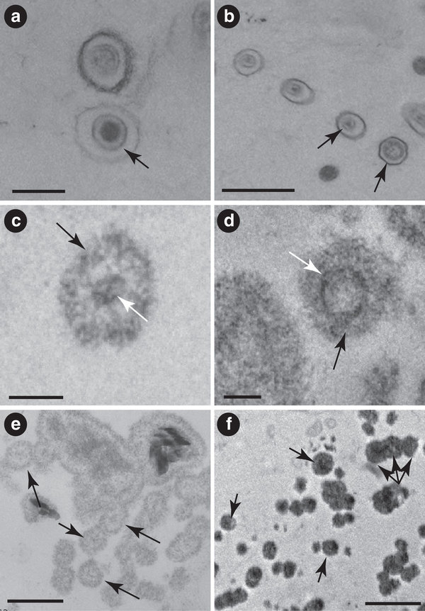
We used the preparation (Diriphagen ® Dr. Heinz Haury Chemical Plant, Munich) because we believed that this preparation met the requirements we just set up. According to Information received, this reparation contains 180-200 different phage strains and thus has a broad spectrum of effectiveness. In addition, it also contains so-called aimed antimicrobics which act against those bacteria that reveal primary phage resistance. We might note here that both the phage components and the added microbics in every ampule are standardized and meet the requirements for biological standardisation as regards phage effect [Penso, G., and Ortali, V., Arch. belges Med. Soc., 1, 1959]. If we mention the two therapeutic components, that is the bacteriphages and aimed (directed) antimicrobics, we are really not fully describing the effects mechanism as such. We have a third factor here. What we are dealing with here is the stimulation of the inherent defenses of the body which are bound to be aroused and which are based on the following: In breeding phages and antimicrobics, the pathogenic microbes used for this purpose give rise to lysates. But these lysates are not eliminated; instead they are also fed into the body. They act like antigens and lead to the formation of antibodies which in turn are specifically directed against the bacteria to which lyntes were added [Glauser, H. A., Med. achr., 13, 420, 1959.]. This reaction requires a latency period of about 8-10 days. The value of this antibody formation is hard to estimate in the individual case. We can get some specific figures on this only if we determine the phagocytosis capability; but this must be done in the clinic. Any new therapy is very often impaired by the fact that we do not employ it until other, more familiar measures have failed. We must admlt that we did not use Deriphagen until we had some patients in whom other preparations had not produced success. This is further by reports from other authors who achieved surprisingly good results with this preparation [Cevey, M., Schweiz. Z. Tuberk. (Swiss Tuberculosis Joural),
15, 34, 1958; Corbelli, G., Bologna Med., 6, 57, 1959; Delacoste, P., Rev. suisse Med., August 1959; Schaefer, W., “Contribution on Epidemic Control” Vol. 3, Hippoprates, Stuttgart, 1948].

Figure 1 shows the result of our treatment. The first column shows the total number of all patients treated; then we have the number of patients cured which abounted to 55.1%; then we cow to those who showed substantial improvement and on the right we have those patients who did not improve as a result of therapy [34.8%].

The author notes, however, that there is a discrepancy between microbiological and clinical results. That is, patients apparently reported a return to healthfulness but this did not coincide with elimination of pathogen, which the author seems to suggest is a consequence of phage- resistant forms not being pathogenic.

The text in the PDF then essentially fades away, though the main text of the paper continues on for two more pages!

Further reading:

Hoeflmayr, J. (1962). Inhalationstherapie mit Bakteriophagen bei therapieresistenten Infektionen [Inhalation Therapy with Bacteriophages for Treatment-Resistant Infections]. Fortschritte der biologischen Aerosol-Forschung in den Jahren 1957–1961 [Advances in Biological Aerosols Research in the Years 1957–1961].  403-409. 1962. (I believe this is the original reference)

Abedon, S. T., Kuhl, S., Blasdel, R., Kutter, E. M. (2011). Phage Treatment of Human Infections. Bacteriophage 1(2): 66-85. [PubMed link] (this article provides further historical context on European use of phage therapy, though note that description of a German tradition in that article is completely lacking and so far as I am aware was unknown to the authors at the time of its writing)