Bacteriophages, Spatial Structure, and the Joys and Limitations of a Swiss Pass

Stephen T. Abedon

Department of Microbiology – The Ohio State University

phage.org – phage-therapy.org – biologyaspoetry.org

(This essay was written while touring Switzerland by train, July, 2014)


Travel can be joyous but also can involve a lot of work. The basic premise of travel is movement, whether specifically from one destination to another or instead something more random. In either case it takes time for you to move from that one place to another. Even the exploration of a smallish country therefore can take enormous amounts of time, since each of numerous legs of your journey will take some amount of time to traverse. You can purchase a Swiss Pass, and explore much of Switzerland over days and even weeks. You’ll see a lot, but you certainly will see far from everything. These temporal delays that are manifest as you travel are one of things that makes traveling difficult, but at the same time this relative slowness can be what makes a journey worthwhile. If you flittered from place to place at the speed of light, never pausing, you would touch upon much more, but your experience would be far different. Indeed, there are qualitative differences between your experiences as you fly, drive, take a train, ride a motorcycle, ride a bike, walk, or indeed not move at all.

Spatial structure is a property of environments in which delays in movement exist. If you can instantaneously and randomly be anywhere, then there is no spatial structure. In microbiology, spatial structure is seen especially under circumstances that do not substantially involve turbulent flow. When you shake a broth-filled flask, one of the consequences of that action is to reduce spatial structure. In terms of interactions between predators and prey, of bacteriophages and bacteria, the result is that any one individual may interact with any other individual with equivalent probability. If you replace any collision between phages and bacteria with the special kind of interaction that is sex, then you have random mating. If you replace any collision with the special kind of interaction that is phage infection of bacteria, then you have random infection. Either case is implicitly a consequence of a lack of spatial structure in the environment.

Spatial structure generally is what happens within environments almost no matter what. You can strive to remove spatial structure, such as via the shaking or stirring of broth, but absent such measures, or indeed if volumes are large enough and mixing slow enough, then some spatial structure nonetheless will be retained. A static microcosm – where the mixing of broth is reduced essentially to zero and therefore where movement is dominated by either motility or diffusion – thus can represent a spatially structured environment. More obvious is the spatial structure that occurs when movement is reduced even further, as is the case with the addition to environments of various thickening agents such as agar.

In phage biology the classic laboratory-observed consequence of spatial structure is the formation of phage plaques, which are clearings within otherwise turbid bacterial cultures, ones that have been spread or poured onto agar plates. The formation of a plaque requires three processes: phage population growth, phage-mediated reduction of bacterial densities, and, crucially, limitations in phage as well as bacterial movement.  Generally a plaque begins with a single plaque forming unit (PFU) which consists of an infective center and in turn can be either an individual phage virion, a clump of phage virions, a phage-infected bacterium, or a clump of bacteria at least one of which is phage infected. This infective center serves as a point source for the outward but nonetheless slow diffusion of phage virions away from their origin. The movement is outward only because the random process of diffusion tends to result in a broadening of the “cloud” of diffusing particles. Because of limitations on the rate of this movement, however, the cloud remains relatively small: the confluent lysis of an entire plate via the growth of a single plaque generally does not occur.

The phage is you. You can start a family and outfit each member of your family with a Swiss Pass. But unless your explorations of the amazing beauty that is Switzerland occurs over extremely long periods, then your and your family’s potential to see all of Switzerland will be relatively limited. This is less true, however, if your family is very large, so large that at least one family member is present to explore each place that may be explored. In this case complete exploration of a discrete area may be achieved. Your ability to explore broader areas nevertheless will be limited at least in part by how long it takes you or your family to get there.

Further reading:

Abedon, S. T., Bartom, E. (2013). Plaques. Brenner’s Encyclopedia of Genetics. Maloy, S., Hughes, K. (eds). Academic Press, pp. 357-357.

Abedon, S. T., Yin, J. (2009). Bacteriophage Plaques: Theory and Analysis. Methods in Molecular Biology 501:161-174. [PubMed link]

Abedon, S. T., Yin, J. (2008). Impact of Spatial Structure on Phage Population Growth. In: Bacteriophage Ecology, Abedon, S. T. (ed), Cambridge University Press, Cambridge, pp. 94-113.

For videos of my explorations of Switzerland, as well as other aspects of my existence, see youtube.com/channel/UCf0uLeBfCToHT3eAoYFmcNA.

crAssphage

A new study led by researchers at San Diego State University has found that more than half the world’s population is host to a newly described virus, named crAssphage, which infects one of the most
common gut bacterial species, Bacteroides. This bacterium is thought to be connected with obesity, diabetes and other gut-related diseases.

Big fleas have little fleas, Upon their backs to bite ’em, And little fleas have lesser fleas, and so, ad infinitum.

One of the many problems farmers of various kinds of legumes need to deal with is the pea aphid, which reproduce incredibly fast and live by sucking the sap out of the plants. However, while they are terrifying parasites of legumes, they have their own yet more horrific parasites, a parasitoid wasp. Below is a really nice close up picture of one doing its thing, here is a video of the act, and here is a brain meltingly horrific video of a dissection of the mummified aftermath 8 days later. Essentially, these wasps deposit their eggs in a pea aphid and the growing larva feeds on it, developing there for about a week, and then consuming the host from the inside out like a Xenomorph. When it’s done, the wasp larva dries the aphid’s cuticle into a papery brittle shell and an adult wasp emerges from the aphid mummy. Legume farmers love these wasps as much as they despise the aphids that destroy their crops, however, when farmers noticed that the wasps didn’t work as effectively on all of aphids infestations, Nancy Moran’s group at the University of Texas in Austin went to work figuring out why. It turns out that all aphids have a primary bacterial endosymbiont living inside their cells, in addition to and much like a mitochondria, and that many have some combination of five other known secondary endosymbionts. Interestingly, two of those other five, Hamiltonella defensa and Serratia symbiotica have been shown to confer varying levels of resistance to the parasitoid wasp, allowing the aphid to survive infection. However, it turns out that there is yet one more layer to this story,

The relationship these endosymbionts have with the aphid, as well as the primary endosymbiont, is hard to classify as they confer a fitness cost in the absence of the wasp but a significant fitness boost when the wasps are around and trying to infect the aphids. At least for H. defensa, the reason why some strains are fully parasitic and provide no protection against the wasps while others are at least plausibly commensal and do provide protection, is a bacterial virus that infects the endosymbiont, even while it is inside the eukaryotic aphid cell. To understand why it will require a bit of knowledge of how some bacteriophages work. Most bacterial viruses, also known as bacteriophages, have a clear dividing line between two strategies. The simplest and most virulent phages will always immediately shut down their host’s metabolism upon infection and replace it with their own. Within a short period of time, generally between 20 and 80 minutes, the phage will have used the host cell to replicate its genome, build new viral particles, packed those particles with the genome and lysed the cell; setting loose 30-3000 new inert infectious particles. These are known as obligately lytic phages. Most phages however, use a mix of this strategy and another one known as lysogeny. These temperate phages will, at the beginning, decide to either virulently infect, producing particles at the total expense of the host, or hide in the host’s genome and inactivate all of its many host lethal genes. Generally it does this by expressing a transcriptional repressor that prevents expression of everything but the repressor, which incidentally protects the host from subsequent infection by related phages. However, some temperate phages will allow for expression of a genomic cassette that will perform some function of benefit to their host – they might as well since they are completely dependent on their host’s wellbeing while in this stage of their life cycle.
It turns out that there is a temperate bacteriophage called APSE, which is common in H. defensa populations in the aphids, that encodes for a cassette of genes that causes H. defensa to attack the wasp larvae with vicious toxins while the phage hides in the genome of its bacterial host. This makes for a really fascinatingly complex system of interdependencies for each of the agents involved. The phage, the bacterial symbiont, and the aphid are all each united in their interdependent need to combat the wasp that kills all three when it succeeds. However, at the same time, both the phage and the bacteria are dependent on the wasp to apply pressure on the aphid to keep them around – otherwise the aphid would cure itself of both creatures that would then be free-loading. Additionally, the wasp the bacteria, and the phage are all completely dependent on the aphid’s sap sucking ability to sustain them, and the aphid is totally dependent on the farmer to continue growing legumes in massive vulnerable monocultures. Furthermore the farmer and the legumes are dependent on the wasp to combat the aphid and largely helpless against the bacteria and the phage. At the same time, despite all of the interlocking incentives toward cooperation, there are also incentives towards each of these agents cheating each other. The farmer has an incentive to ‘cheat’ and save money by neglecting to buy aphid mummies every so often, because they still benefit from the fitness cost caused by the aphid not rejecting the bacteria or the bacteria rejecting the phage. Similarly, the aphid has an incentive to cheat both the bacteria and the phage to cure itself of them and bet on the farmer not buying aphid mummies full of wasps that year. The bacteria also has an incentive to cheat the aphid by curing itself of the phage, and also bet on the farmer not buying mummies that year itself.
What I love most about this story is that complex series of interdependent yet competing evolutionary interests, which forms as an emergent property of the Siphonaptera – this blog’s namesake,

Big fleas have little fleas, Upon their backs to bite ’em, And little fleas have lesser fleas, and so, ad infinitum. And the great fleas, themselves, in turn Have greater fleas to go on; While these again have greater still, And greater still, and so on.

It is much like another wonderful paper, where a woman’s eye is a big flea bitten the smaller flea of an Acanthamoeba polyphaga parasite,  which is in turn bitten by its Mimiviridae (Lentille virus), which is bitten by its virophage (Sputnik 2), which is itself in a sense bitten by its mobile genetic elements. We have a situation where the farmer is a big flea bitten by their legume, which is bitten by its aphid, which is then bitten by both its parasitoid wasp and its secondary endosymbiont, which is in turn then bitten by its temperate bacteriophage.

This post is deeply indebted to one made on Moselio Schaechter’s excellent blog Small Things Considered, which is slightly more technical and no doubt more clearly written.

Bacteriophages encode factors required for protection in a symbiotic mutualism

Oliver KM, Degnan PH, et al. Published 2009 in Science doi: 10.1126/science.1174463  [REQUIRES FREE REGISTRATION]

Bacteriophages are known to carry key virulence factors for pathogenic bacteria, but their roles in symbiotic bacteria are less well understood. The heritable symbiont Hamiltonella defensa protects the aphid Acyrthosiphon pisum from attack by the parasitoid Aphidius ervi by killing developing wasp larvae. In a controlled genetic background, we show that a toxin-encoding bacteriophage is required to produce the protective phenotype. Phage loss occurs repeatedly in laboratory-held H. defensa–infected aphid clonal lines, resulting in increased susceptibility to parasitism in each instance. Our results show that these mobile genetic elements can endow a bacterial symbiont with benefits that extend to the animal host. Thus, phages vector ecologically important traits, such as defense against parasitoids, within and among symbiont and animal host lineages.

The players in a mutualistic symbiosis: insects, bacteria, viruses, and virulence genes.

Moran NA, Degnan PH, et al. Published 2005 in PNAS USA, doi:10.1073/pnas.0507029102

Aphids maintain mutualistic symbioses involving consortia of coinherited organisms. All possess a primary endosymbiont, Buchnera, which compensates for dietary deficiencies; many also contain secondary symbionts, such as Hamiltonella defensa, which confers defense against natural enemies. Genome sequences of uncultivable secondary symbionts have been refractory to analysis due to the difficulties of isolating adequate DNA samples. By amplifying DNA from hemolymph of infected pea aphids, we obtained a set of genomic sequences of H. defensa and an associated bacteriophage. H. defensa harbors two type III secretion systems, related to those that mediate host cell entry by enteric pathogens. The phage, called APSE-2, is a close relative of the previously sequenced APSE-1 but contains intact homologs of the gene encoding cytolethal distending toxin (cdtB), which interrupts the eukaryotic cell cycle and which is known from a variety of mammalian pathogens. The cdtB homolog is highly expressed, and its genomic position corresponds to that of a homolog of stx (encoding Shiga-toxin) within APSE-1. APSE-2 genomes were consistently abundant in infected pea aphids, and related phages were found in all tested isolates of H. defensa, from numerous insect species. Based on their ubiquity and abundance, these phages appear to be an obligate component of the H. defensa life cycle. We propose that, in these mutualistic symbionts, phage-borne toxin genes provide defense to the aphid host and are a basis for the observed protection against eukaryotic parasites.

It Sometimes Rains Nonsense After Hurricanes

Today I want to talk about a paper that looks incredibly cool in a whole bunch of different ways in the abstract, introduction, and discussion but for whom much of that awesomeness falls apart under closer inspection of the results, methods, and context.  In it, the authors report their results having flown in NASA’s venerable old DC-8 across the US and down the west coast as well as through a couple of hurricanes with a filter designed to capture bacteria sized particles hanging out the side.  They then took the filter and analyzed it with fluorescent dyes and microscopes as well as genomically to see what was there.  In their paper they appear to arrive at five different major conclusions: that viable bacterial cells represented on average around 20% of the total particles in the 0.25- to 1-μm diameter range; that 60 to 100% of the 1.5 × 10⁵ cells m−3 they saw were viable; that bacteria are at least two orders of magnitude more abundant than fungal cells in the troposphere; and that fecal coliforms represent a significant amount of the microbiota of hurricanes after landfall. Additionally, what has made the most splash though, is their speculation that because some of the taxa they determined were present by small subunit rRNA sequencing had been shown to metabolize oxalic acid, a major chemical component of clouds, it was plausible that there was active bacterial metabolism happening in the clouds they analyzed.  Unfortunately, despite the journal it is published in and glowing praise from excellent blogs like Not Exactly Rocket ScienceClimate CentralWiredMetafilter, and The Scientist, the speculation is pretty foolish and each of these conclusions is either inherently false, actively misleading, or very difficult to support with their data.

SnoopyWhat I’d love to do with the Authors’ platform


Here is the paper:

Microbiome of the upper troposphere: Species composition and prevalence, effects of tropical storms, and atmospheric implications

N DeLeon-Rodriguez, TL Lathem, LM Rodriguez-R, et al. Published 2013 in PNAS. doi: 10.1073/pnas.1212089110
The composition and prevalence of microorganisms in the middle-to-upper troposphere (8–15 km altitude) and their role in aerosol-cloud-precipitation interactions represent important, unresolved questions for biological and atmospheric science. In particular, airborne microorganisms above the oceans remain essentially uncharacterized, as most work to date is restricted to samples taken near the Earth’s surface. Here we report on the microbiome of low- and high-altitude air masses sampled onboard the National Aeronautics and Space Administration DC-8 platform during the 2010 Genesis and Rapid Intensification Processes campaign in the Caribbean Sea. The samples were collected in cloudy and cloud-free air masses before, during, and after two major tropical hurricanes, Earl and Karl. Quantitative PCR and microscopy revealed that viable bacterial cells represented on average around 20% of the total particles in the 0.25- to 1-μm diameter range and were at least an order of magnitude more abundant than fungal cells, suggesting that bacteria represent an important and underestimated fraction of micrometer-sized atmospheric aerosols. The samples from the two hurricanes were characterized by significantly different bacterial communities, revealing that hurricanes aerosolize a large amount of new cells. Nonetheless, 17 bacterial taxa, including taxa that are known to use C1–C4 carbon compounds present in the atmosphere, were found in all samples, indicating that these organisms possess traits that allow survival in the troposphere. The findings presented here suggest that the microbiome is a dynamic and underappreciated aspect of the upper troposphere with potentially important impacts on the hydrological cycle, clouds, and climate.

I will start with the most sensational aspect of the paper speculated about by the authors, their assertion that their results indicate it is plausible there is active bacterial life growing in clouds in the troposphere.  While the idea that clouds are themselves life forms, ice seeded by cells and cells fed by oxalic acid attracted by the ice, which the authors go to great lengths to speculate on, is very attractive they neglect to mention the temperature readings taken during the flights anywhere in the paper or even supplementary information, which should have pretty much immediately dismissed all of it.  The troposphere that the authors were sampling is typically between -50 and -70°C, which approaches the kinds of temperatures I use in my lab to keep bacterial cells in immortal suspended animation.   Indeed, even the most extreme psychrophiles don’t grow much below -12°C and even then only really in the presence of a large amount of salt that helps them keep the water they’re in from freezing.  The bacteria that they saw may not have been dead exactly when they were sampled, but they certainly weren’t living, which brings us to their next sexy conclusion.

The authors claim that viable bacterial cells represented on average around 20% of the total particles in the 0.25- to 1-μm diameter range and that 60 to 100% of the 1.5 × 10⁵ cells m−3 they saw were viable. This implies that even if the bacteria involved were not actively living, many of them were at least only mostly dead, in a Princess Bride sense. Unlike ordinary humans, bacteria can generally quite happily be frozen in place and exist suspended indefinitely, coming back to life should they thaw in a favorable situation. To determine just how dead the bacteria they were looking at were, the authors used a set of fluorescent dyes sold by Invitrogen that stains DNA green if it is surrounded by an intact membrane, or red if not, on particles they liberated from their filters.   However, while the promotional materiel for the various kinds of kits they could have used from their description talk a big game about determining viability, that is not something they can do, at least not for these authors.  All that those dyes can measure is whether cells are intact or not, which in some well characterized kinds of systems can be an excellent proxy for whether or not they are viable, but the authors have no idea if it is for them and it is in fact really profoundly unlikely.  Even setting aside the likelihood that a significant portion of what they were staining was not DNA to begin with, the upper atmosphere is an incredibly hostile place that one would expect to leave cells a lot more than just mostly dead, even while leaving them intact.  Ultraviolet radiation from the sun would kink the DNA of anything up there causing the same kinds of damage as a sunburn, while the dry conditions should dessicate all but the hardiest spores.  This is not to say that it isn’t plausible that there truly are viable bacteria up there, but it is to say that the authors do not measure viability in a remotely meaningful way and cannot really contribute to our understanding of how viable exactly those bacteria might be.

Picture1

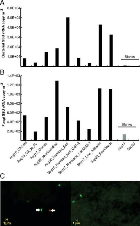
Fig. 1. Quantification of bacterial and fungal cells in samples from high altitudes in the atmosphere. Concentration of bacterial (A) and fungal (B) cells based on qPCR analysis of SSU rRNA gene copies in the samples. Note that samples are ordered by the collection time on the x axis except for blank samples, which are shown at the rightmost part of the graphs in light gray. (C) Live/dead microscopy image of two samples from the California coast and transit flights. Green-stained cells represent cells with viable/intact membrane (e.g., cell indicated by left arrow), and red/yellow-stained cells represent cells with a damaged membrane (e.g., cell indicated by right arrow). Credit: (DeLeon-Rodriguez et al., 2013)

The authors also conclude based on the data shown above that bacteria are at least two orders of magnitude more abundant than fungal cells in the troposphere, and that is indeed what their data plausibly shows. This is to say that even though all of the qPCR data they are relying on is very weak having massive amounts of contamination in their control blanks (just look at the scale), they are blind to that contamination having failed to sequence it, they have no replicates to do statistics with, and they get results that are inconsistent with their other data by two orders of magnitude – they still make a decent case that they see less fungal DNA than bacterial DNA in their filters and that that means fewer fungal cells than bacterial cells. They do however also neglect to mention anywhere in the paper that fungi could be reasonably expected to be less abundant in the late summer when they were measuring, particularly in relation to the early Spring or to a lesser extent mid Fall when fungi tend to sporulate.

Picture2

Fig. 3. Habitat of origin of the SSU rRNA gene sequences recovered in the GRIP samples. Sequences were assigned to a habitat (see key) based on the source of isolation of their best match in the GreenGenes database. The graph represents the relative abundance of each habitat (vertical axis) for each sample (x axis). Numbers on the top denote the fraction of sequences that were assignable to a habitat for each sample. Credit: (DeLeon-Rodriguez et al., 2013)

The last major finding of this paper, that fecal coliforms represent a significant amount of the microbiota of hurricanes after they make landfall is a really cool one, but if the authors can demonstrate that this is the case, they don’t do it convincingly in this paper.  Even setting aside the question of how viable the fecal coliforms they saw were, there is still the very tricky question of the DNA contamination they saw.  While I’m sure we can trust that this wasn’t contamination with lab strains from say the autoclave they used for sterilization, which they speculated might be a source in the paper as that would show up very obviously as having little to no diversity, the amount of coliforms they saw is still well within the levels of contamination they know they have.  This could all plausibly come from say a livestock operation next to the airport they left from and returned to or some other artifact of the post landfall expeditions they made.

Flight trajectories

Figure S1.  Flight trajectory maps.  (A) Flights conducted in the west coast (red) and across the USA (blue).  (B) Flights conducted in the area of the Caribbean Sea and the mid-western Atlantic Ocean. The route of each flight is color-coded (see figure key). The trajectory of Hurricane Earl and Karl are colored-coded based on the intensity of the hurricane at each time point (scale bar). Credit: (DeLeon-Rodriguez et al., 2013)

I think it is a particular shame that this research has been sexed up as a microbiota paper that it is not, or at least is not yet, because it still looks like a very interesting climate and meteorological paper.  They still are able to convincingly show that that intact bacterial cells in the atmosphere, and particularly within hurricanes, are at least within the same order of magnitude as particles from non-biological origins – this is a really cool and, at least as far as I can tell, novel finding outside of dust from the Sahara.    Even if they say silly things about how alive those cells are, have nothing really they could say about how only mostly dead those cells could be, and are limited in what they could say about what kinds of cells they see, that doesn’t mean they couldn’t use stronger techniques in the future.  Really to say the kinds of things they want to say, the authors would absolutely need to use culture dependent methods to look at their filters.  You just cannot have anything meaningful to say about a bacteria’s ability to grow without actually growing some bacteria.  Now, culture dependent methods do have their weaknesses, namely that using them, we only seem to be able to grow about 1% of the bacteria present on Earth, but with the kinds of huge numbers of bacteria the authors are throwing around that shouldn’t scare them.  It would also help them to do something I would find really cool: look for viruses.

If they’re actually serious about the contention that there are critters actually actively alive up there, there would be nothing stopping bacteriophages from infecting them, and showing their presence would go a long way to demonstrating that there is ecology going on in clouds. The idea that hurricanes could spread viable cells through the troposphere would go a long way towards explaining why we routinely do things like find in American belly buttons a bacteria that had previously only been found once in Japanese soil; finding viable (by which I strictly mean culturable) bacteriophages in the troposphere would go a long way towards explaining why we see the same thing with them. Now all I need to do is convince the NSF to let me look!

See also a letter to PNAS and its response in the next issue:

Inadequate methods and questionable conclusions in atmospheric life study (Smith & Griffin, 2013)

and

Reply to Smith and Griffin: Methods, air flows, and conclusions are robust in the DeLeon-Rodriguez et al. study (DeLeon-Rodriguez et al., 2013)


The origin of virulence, and why its important.

Virulence is an abstraction of the harm caused to hosts by a pathogen, and explaining the paradox of virulence has been an active field of study in evolution for a while. In general the harm caused to the hosts of pathogens is not great for the pathogen, after all, why hurt or lose a useful host? However, in studying the abstraction with basic research, we’ve found that virulence is almost always is part of helping the pathogen find a new host. Thus the generalized answer to the paradox is that so long as the harm to the host causes the parasite to spread effectively enough, it doesn’t really matter how much harm is caused to the host – as the parasite will have already found new hosts to spread from. At the same time, helpful bacteria don’t have nearly the same need to spread as pathogenic ones, as they keep their hosts happy and alive and can stick around for longer.

Here I’ll introduce two papers demonstrating this model and try to convince you of how important it is.

The spectrum between virulence and mutualism can be seen as a trade off between two strategies, as well as of course often a mix between the two. A critter existing in community with another one can care little for its host and work to be as infectious as possible at the host’s expense, thus increasing virulence. In this strategy it doesn’t matter so much that the host becomes quickly unsuitable so long as the parasite has already found replacement hosts sneezed on, or transmitted to, by the time that happens. Or it can do the opposite and try its best to reduce impact on the host, spread infectious particles slowly or even not at all, and thus not need to spread too quickly because it will last a while in each host. Most of the critters that live in our guts and on our skin are at this end of the spectrum, and have become so adept at not messing up their host as to actually benefit us in some way. On the other end of the spectrum are parasitoids. These are the parasites that not only destroy their host in their race to infect as many more hosts as possible, but spend the majority of their life cycle doing so and ultimately sterilize or kill, and sometimes consume the host in the process. The Xenomorphs from the movie Alien are a beautiful example of a bunch of these sorts of parasitiod strategies, each inspired by real terrifying stuff in nature. This might all seem uselessly theoretical, but the implications it has for public health are really cool.

Before the advent of antibiotics, we lived with Staphylococcus aureus strains on our skin that existed in a complex mixture of commensal and virulent strategies, but antibiotics suddenly applied very strong selective pressure against any vaguely virulent strategy. Thus, following the model, the observed sudden decrease in both virulence and transmissibility of virulent strains makes a lot of sense. However, the sudden increase in both virulence and transmissibility of virulent strains that we’ve seen in multi-drug resistant (MRSA) strains also makes sense. Indeed, if you look back far enough in the literature all of the crazy new and terrible virulence factors we are now seeing in MRSA strains all existed before the 1930s. For example, while the pyomyositis and necrotizing pneumonia we are now seeing is commonly associated with poverty, tropical climates and HIV, ie: things which didn’t get much attention prior to 1935, it was described. (At lest with this source you’ll need to wade your way past the kinds of phrases that start with “Africans are not different from any other humans, however, …” to page 1214) Until recently it would not be terribly remarkable, being easily addressed with a simple round of I.V. antibiotics.  Additionally, the PVL toxin which that first paper describes as now being found in pneumonia was initially discovered by Van deVelde in 1894 and was named after Sir Philip Noel Panton and Francis Valentine when they associated it with soft tissue infections in 1932. All of this makes logical sense anyhow, the mechanisms of antibiotic resistance are not associated with pathogenesis.

Timing of transmission and the evolution of virulence of an insect virus.

JC de Roode, AJ Yates, & S Altizer. Published 2002 in Proc. R. Soc. Lond. B doi:10.1098/rspb.2002.1976

We used the nuclear polyhedrosis virus of the gypsy moth, Lymantria dispar, to investigate whether the timing of transmission influences the evolution of virulence. In theory, early transmission should favour rapid replication and increase virulence, while late transmission should favour slower replication and reduce virulence. We tested this prediction by subjecting one set of 10 virus lineages to early transmission (Early viruses) and another set to late transmission (Late viruses). Each lineage of virus underwent nine cycles of transmission. Virulence assays on these lineages indicated that viruses transmitted early were significantly more lethal than those transmitted late. Increased exploitation of the host appears to come at a cost, however. While Early viruses initially produced more progeny, Late viruses were ultimately more productive over the entire duration of the infection. These results illustrate fitness trade-offs associated with the evolution of virulence and indicate that milder viruses can obtain a numerical advantage when mild and harmful strains tend to infect separate hosts.

Virulence-transmission trade-offs and population divergence in virulence in a naturally occurring butterfly parasite (PDF).

VS Cooper, MH Reiskind, et al. Published 2002 in PNAS doi:10.1073/pnas.0710909105

Why do parasites harm their hosts? Conventional wisdom holds that because parasites depend on their hosts for survival and transmission, they should evolve to become benign, yet many parasites cause harm. Theory predicts that parasites could evolve virulence (i.e., parasite-induced reductions in host fitness) by balancing the transmission benefits of parasite replication with the costs of host death. This idea has led researchers to predict how human interventions—such as vaccines—may alter virulence evolution, yet empirical support is critically lacking. We studied a protozoan parasite of monarch butterflies and found that higher levels of within-host replication resulted in both higher virulence and greater transmission, thus lending support to the idea that selection for parasite transmission can favor parasite genotypes that cause substantial harm. Parasite fitness was maximized at an intermediate level of parasite replication, beyond which the cost of increased host mortality outweighed the benefit of increased transmission. A separate experiment confirmed genetic relationships between parasite replication and virulence, and showed that parasite genotypes from two monarch populations caused different virulence. These results show that selection on parasite transmission can explain why parasites harm their hosts, and suggest that constraints imposed by host ecology can lead to population divergence in parasite virulence.