Phages & Flamingos

For my first post on this blog, I wanted to share the following paper with you, The virus’s tooth: cyanophages affect an African flamingo population in a bottom-up cascade (see below). It captured my attention after Dr. Brian Jones visited my lab earlier in 2014 where he gave a lecture on flamingos in Kenya (Lesser Flamingo, Phoeniconaias minor). As an extremophile specialist he had been invited to a workshop organized by the Kenyan government to find out why the flamingos have disappeared from Lake Nakuru, a local alkaline lake. According to Brian, many theories were offered up during the workshop, but none of them with sufficient evidence, mainly because of a lack of long-term monitoring of the lakes’ ecosystem.

The following paper presents a possible explanation: Phages caused it! The researchers hypothesize that cyanophages are at the root of a bottom-up cascade causing the flamingo’s main food source, the cyanobacterium Arthrospira fusiformis, to be broken down causing massive drops in flamingo numbers. 

A question of where did the flamingos go, was partly answered accidently at a sampling expedition of my lab, the Centre for Microbial Ecology and Genomics (CMEG, University of Pretoria). Each year a bunch of researchers of CMEG and collaborators make a trip to the Namib Desert to investigate the local arid ecosystems. When driving to the closest town, Walvis Bay, about a 90 minute away located at the Atlantic Ocean, many people stop at the actual bay to watch huge gatherings of the Lesser Flamingo. Sadly, we have no records of how many years the flamingos have been gathering there and if there stay there year-round or not.


The virus’s tooth: cyanophages affect an African flamingo population in a bottom-up cascade

Link to the article

ABSTRACT

Trophic cascade effects occur when a food web is disrupted by loss or significant reduction of one or more of its members. In East African Rift Valley lakes, the Lesser Flamingo is on top of a short food chain. At irregular intervals, the dominance of their most important food source, the cyanobacterium Arthrospira fusiformis, is interrupted. Bacteriophages are known as potentially controlling photoautotrophic bacterioplankton. In Lake Nakuru (Kenya), we found the highest abundance of suspended viruses ever recorded in a natural aquatic system. We document that cyanophage infection and the related breakdown of A. fusiformis biomass led to a dramatic reduction in flamingo abundance. This documents that virus infection at the very base of a food chain can affect, in a bottom-up cascade, the distribution of end consumers. We anticipate this as an important example for virus-mediated cascading effects, potentially occurring also in various other aquatic food webs. 


Phage Therapy Case Study from 1936

Stephen T. Abedon

Department of Microbiology – The Ohio State University

phage.org – phage-therapy.org – biologyaspoetry.org


This article can’t be found via a PubMed search but can be found here: jama.jamanetwork.com/article.aspx?articleid=1156439. It is not free, but most of it can be found on that page. The reference is Morrison, S., Gardner, R.E. (1936). The Treatment of a Lung Abscess due to Bacillus coli with a Lytic Filtrate. JAMA 107(1):33-34. It is a fascinating account because it walks you through the case in some detail plus presents both efficacy and side effects, neither of which can be unquestionably attributed to the phage itself since the formulation used was not purified. Still, pretty amazing stuff, and I quote:

N, S., a woman, aged 22, who had previously been in excellent health, suddenly experienced a severe diffuse abdominal pain, Aug. 5, 1934… On the third day the patient’s condition became critical and she was rushed to the Chambersburg (Pa.) Hospital, where an emergency operation was performed by Dr. L. H. Seaton. When the abdomen was opened a gangrenous appendix with generalized peritonitis was disclosed. The remainder of the appendix was removed and drains were inserted…

[Approximately one month later,] after an excruciating pain, examination disclosed massive collapse of the left lung. During the subsequent few days slight signs of partial return of pulmonary function were observed, but relapse followed. Clinical and x-ray signs of effusion developed. Aspiration was performed September 12 and 500 cc. of very heavy purulent material with a foul and typical colon odor was obtained. A culture of the pus at this time yielded only Bacillus coli. Three days later, because the material was too thick to be aspirated, rib resection was done with a virtual gush of pus. A bronchial fistula developed shortly after the rib resection and the patient was expectorat¬ ing the same kind of material as that which drained from the resection wound. The appearance of the area around the resection opening was necrotic and “mossy” and failed to show any improvement on local irrigations with 1,000 cc. of saline solution twice a day. Digital examination through the resection wound disclosed many walled off abscesses surrounded by necrotic tissue. In view of the hectic fever and the general condition, which indicated toxic absorption, an especially resistant abscess which failed to open was incised by an approach between the ribs just above the rib resection. A drain was inserted and in a few days healing took place.

A second sample of pus was collected at this time (September 16) and another pure culture of colon bacillus isolated which was fairly readily lysed by a bacteriophage that was active against various strains of B. coli isolated from other sources.

After a cutaneous test September 20 of 0.1 cc. of the lytic filtrate twelve hours previously had given little or no reaction, and after irrigating the chest with 1 liter of physiologic solution of sodium chloride, 1 ounce (30 cc.) of the phage was instilled and allowed to remain for two hours. This was followed saline irrigation and the wound covered by a dressing saturated with the bacteriophage. The following day the observation was made that the discharge had become thin and watery and had lost its offensive character for the first time since the resection was done five days before, even though saline irrigations had been administered twice daily during this five day period. A second and equally remarkable change had occurred at the resection wound itself, where the mossy, necrotic character was entirely changed to a clean, fresh, healthy appearing incision.

Since the first use of bacteriophage had given such excellent results, a second application seemed indicated, and therefore the procedure was repeated. However, within ten minutes a violent generalized rose-colored urticaria appeared and the patient complained of nausea and vomited. The bacteriophage was drained immediately and the chest irrigated with large quantities of saline solution. Epinephrine was administered…

After such a marked allergic reaction to the bacteriophage had occurred it was decided to discontinue bacteriophage instillations and continue only with saline irrigations and external dressings saturated with bacteriophage. The dressings of bacteriophage were continued for a week along with irrigations of physiologic solution of sodium chloride. Throughout this period the resection wound maintained its healthy normal appearance and the discharge remained clear, watery and nonodorous. The temperature reached 102.2 F. each day for the thirteen days prior to the urticarial reaction. On that day the reading was 103.2 F. after the reaction. After this reaction the temperature did not go above 102.2 F.

The patient’s general condition was remarkably improved and within six weeks she was able to leave the hospital. The appendiceal wound had healed but the fever, less hectic in type, continued as well as the thin nonodorous drainage. At home the fever gradually subsided as well as the drainage, and heal¬ ing was practically complete toward the end of December.

Whether the bacteriophage acted as a specific or indirectly as a Synergist to antibody formation cannot be stated.

Thus, no proof of explicitly phage-mediated efficacy, no proof that the condition would not have spontaneously reversed on its own, and no controls, but instead a remarkable result, with an indication as well of reason for caution regarding potential immunological reactions perhaps associated with the lack of formulation purification. Interesting indeed!

Bacteriophage-based synthetic biology for the study of infectious diseases

An interesting new short paper out of Tim Lu’s synthetic biology lab showcasing what bacteriophage can bring to the table,

Bacteriophage-based synthetic biology for the study of infectious diseases

Robert J Citorik, Mark Mimee, and Timothy K Lu Published 2014 in Curr. Opin Microbiol. DOI: 10.1016/j.mib.2014.05.022

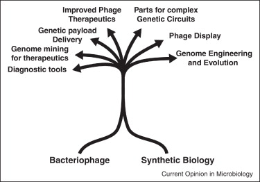
Since their discovery, bacteriophages have contributed enormously to our understanding of molecular biology as model systems. Furthermore, bacteriophages have provided many tools that have advanced the fields of genetic engineering and synthetic biology. Here, we discuss bacteriophage-based technologies and their application to the study of infectious diseases. New strategies for engineering genomes have the potential to accelerate the design of novel phages as therapies, diagnostics, and tools. Though almost a century has elapsed since their discovery, bacteriophages continue to have a major impact on modern biological sciences, especially with the growth of multidrug-resistant bacteria and interest in the microbiome.

•Multidrug-resistant infections have sparked a renewed interest in bacteriophages.
•Synthetic biology has enabled technologies for next-generation phage engineering.
•Engineered phages and derived parts constitute a new antimicrobial paradigm.
•Bacteriophage-based reporters permit detection of specific pathogens.
•Components from phage form a core set of parts in the synthetic biology toolbox.

Bacteriophage - Synthetic Biology

A Quote or Two from Hoeflmayr (1963): “Inhalation Therapy Using Bacteriophages in Therapy-Resistant Infections”

Stephen T. Abedon

Department of Microbiology – The Ohio State University

phage.org – phage-therapy.org – biologyaspoetry.org


I just came across this “report”, which can be found here:

http://www.dtic.mil/cgi-bin/GetTRDoc?Location=U-2&docGetTRDoc.pdf&AD=AD0837021

This dates from 1963, I believe as a translation, and the full citation is “Inhalation Therapy Using Bacteriophages in Therapy-Resistant Infections”. Fortschritte der Biologischen Aerosol-Forschung-Jahren 1957-1961 (Progress of the Biological Aerosol Research-Years 1957-1961), pp. 403-409. See under “Further reading” for what presumably is the original and/or complete citation.

At any rate, this paper/chapter/publication/translation/report has some interesting passages.

In view of the growing resistance against antibiotics, it is vitally important that we try to find ways to counteract this development. … Farsighted clinicians warned us as long as 10 years ago, when we were still students, that we should not hastily treat any little infection with penicillin.
If we should discover any-new possibilities for treating infections, then we should look at these possibilities only from the angle that such a therapy would have to preclude the formation of resistance as much as possible, therapy with bacteriophages fills the requirement. The fact that this therapy has so far met with skepticism is due to the results which, until a few years ago, did not ome up to expectations
[Schaefer, W., “Contribution on Epidemic Control” Vol. 3, Hippocrates, Stuttgart, 1948]. If we try to track down the reason for the failure of the earlier bacteriophage therapy, we will find that this was mostly due to the biological properties of the phages.
Now it is important to know that the bacteriophage has high specificity. Therefore, therapy can be effective
only If the administered phage encounters its homologous bacterium.
The disadvantage of our earlier phage preparations was to be found not only in the inadequate breeding methods but above all in the fact that only about 1-2 phage strains were available. If we consider the large number of pathogenic bacteria strains, which play a role even in a very simple infection or which at least at times might play a role, then we would have to set up two requirements. First of all, in order to have a wide range of effectiveness, such a therapeutic substance would have to contain a large number of various phage strains. Second, it is necessary that phages which would come into consideration for therapy should have sufficient virulence with regard to pathogenic viruses.
We used the preparation (Diriphagen ® Dr. Heinz Haury Chemical Plant, Munich) because we believed that this preparation met the requirements we just set up. According to Information received, this reparation contains 180-200 different phage strains and thus has a broad spectrum of effectiveness. In addition, it also contains so-called aimed antimicrobics which act against those bacteria that reveal primary phage resistance. We might note here that both the phage components and the added microbics in every ampule are standardized and meet the requirements for biological standardisation as regards phage effect [Penso, G., and Ortali, V., Arch. belges Med. Soc., 1, 1959]. If we mention the two therapeutic components, that is the bacteriphages and aimed (directed) antimicrobics, we are really not fully describing the effects mechanism as such. We have a third factor here. What we are dealing with here is the stimulation of the inherent defenses of the body which are bound to be aroused and which are based on the following: In breeding phages and antimicrobics, the pathogenic microbes used for this purpose give rise to lysates. But these lysates are not eliminated; instead they are also fed into the body. They act like antigens and lead to the formation of antibodies which in turn are specifically directed against the bacteria to which lyntes were added [Glauser, H. A., Med. achr., 13, 420, 1959.]. This reaction requires a latency period of about 8-10 days. The value of this antibody formation is hard to estimate in the individual case. We can get some specific figures on this only if we determine the phagocytosis capability; but this must be done in the clinic. Any new therapy is very often impaired by the fact that we do not employ it until other, more familiar measures have failed. We must admlt that we did not use Deriphagen until we had some patients in whom other preparations had not produced success. This is further by reports from other authors who achieved surprisingly good results with this preparation [Cevey, M., Schweiz. Z. Tuberk. (Swiss Tuberculosis Joural),
15, 34, 1958; Corbelli, G., Bologna Med., 6, 57, 1959; Delacoste, P., Rev. suisse Med., August 1959; Schaefer, W., “Contribution on Epidemic Control” Vol. 3, Hippoprates, Stuttgart, 1948].

Figure 1 shows the result of our treatment. The first column shows the total number of all patients treated; then we have the number of patients cured which abounted to 55.1%; then we cow to those who showed substantial improvement and on the right we have those patients who did not improve as a result of therapy [34.8%].

The author notes, however, that there is a discrepancy between microbiological and clinical results. That is, patients apparently reported a return to healthfulness but this did not coincide with elimination of pathogen, which the author seems to suggest is a consequence of phage- resistant forms not being pathogenic.

The text in the PDF then essentially fades away, though the main text of the paper continues on for two more pages!

Further reading:

Hoeflmayr, J. (1962). Inhalationstherapie mit Bakteriophagen bei therapieresistenten Infektionen [Inhalation Therapy with Bacteriophages for Treatment-Resistant Infections]. Fortschritte der biologischen Aerosol-Forschung in den Jahren 1957–1961 [Advances in Biological Aerosols Research in the Years 1957–1961].  403-409. 1962. (I believe this is the original reference)

Abedon, S. T., Kuhl, S., Blasdel, R., Kutter, E. M. (2011). Phage Treatment of Human Infections. Bacteriophage 1(2): 66-85. [PubMed link] (this article provides further historical context on European use of phage therapy, though note that description of a German tradition in that article is completely lacking and so far as I am aware was unknown to the authors at the time of its writing)
Big fleas have little fleas, Upon their backs to bite ’em, And little fleas have lesser fleas, and so, ad infinitum.

One of the many problems farmers of various kinds of legumes need to deal with is the pea aphid, which reproduce incredibly fast and live by sucking the sap out of the plants. However, while they are terrifying parasites of legumes, they have their own yet more horrific parasites, a parasitoid wasp. Below is a really nice close up picture of one doing its thing, here is a video of the act, and here is a brain meltingly horrific video of a dissection of the mummified aftermath 8 days later. Essentially, these wasps deposit their eggs in a pea aphid and the growing larva feeds on it, developing there for about a week, and then consuming the host from the inside out like a Xenomorph. When it’s done, the wasp larva dries the aphid’s cuticle into a papery brittle shell and an adult wasp emerges from the aphid mummy. Legume farmers love these wasps as much as they despise the aphids that destroy their crops, however, when farmers noticed that the wasps didn’t work as effectively on all of aphids infestations, Nancy Moran’s group at the University of Texas in Austin went to work figuring out why. It turns out that all aphids have a primary bacterial endosymbiont living inside their cells, in addition to and much like a mitochondria, and that many have some combination of five other known secondary endosymbionts. Interestingly, two of those other five, Hamiltonella defensa and Serratia symbiotica have been shown to confer varying levels of resistance to the parasitoid wasp, allowing the aphid to survive infection. However, it turns out that there is yet one more layer to this story,

The relationship these endosymbionts have with the aphid, as well as the primary endosymbiont, is hard to classify as they confer a fitness cost in the absence of the wasp but a significant fitness boost when the wasps are around and trying to infect the aphids. At least for H. defensa, the reason why some strains are fully parasitic and provide no protection against the wasps while others are at least plausibly commensal and do provide protection, is a bacterial virus that infects the endosymbiont, even while it is inside the eukaryotic aphid cell. To understand why it will require a bit of knowledge of how some bacteriophages work. Most bacterial viruses, also known as bacteriophages, have a clear dividing line between two strategies. The simplest and most virulent phages will always immediately shut down their host’s metabolism upon infection and replace it with their own. Within a short period of time, generally between 20 and 80 minutes, the phage will have used the host cell to replicate its genome, build new viral particles, packed those particles with the genome and lysed the cell; setting loose 30-3000 new inert infectious particles. These are known as obligately lytic phages. Most phages however, use a mix of this strategy and another one known as lysogeny. These temperate phages will, at the beginning, decide to either virulently infect, producing particles at the total expense of the host, or hide in the host’s genome and inactivate all of its many host lethal genes. Generally it does this by expressing a transcriptional repressor that prevents expression of everything but the repressor, which incidentally protects the host from subsequent infection by related phages. However, some temperate phages will allow for expression of a genomic cassette that will perform some function of benefit to their host – they might as well since they are completely dependent on their host’s wellbeing while in this stage of their life cycle.
It turns out that there is a temperate bacteriophage called APSE, which is common in H. defensa populations in the aphids, that encodes for a cassette of genes that causes H. defensa to attack the wasp larvae with vicious toxins while the phage hides in the genome of its bacterial host. This makes for a really fascinatingly complex system of interdependencies for each of the agents involved. The phage, the bacterial symbiont, and the aphid are all each united in their interdependent need to combat the wasp that kills all three when it succeeds. However, at the same time, both the phage and the bacteria are dependent on the wasp to apply pressure on the aphid to keep them around – otherwise the aphid would cure itself of both creatures that would then be free-loading. Additionally, the wasp the bacteria, and the phage are all completely dependent on the aphid’s sap sucking ability to sustain them, and the aphid is totally dependent on the farmer to continue growing legumes in massive vulnerable monocultures. Furthermore the farmer and the legumes are dependent on the wasp to combat the aphid and largely helpless against the bacteria and the phage. At the same time, despite all of the interlocking incentives toward cooperation, there are also incentives towards each of these agents cheating each other. The farmer has an incentive to ‘cheat’ and save money by neglecting to buy aphid mummies every so often, because they still benefit from the fitness cost caused by the aphid not rejecting the bacteria or the bacteria rejecting the phage. Similarly, the aphid has an incentive to cheat both the bacteria and the phage to cure itself of them and bet on the farmer not buying aphid mummies full of wasps that year. The bacteria also has an incentive to cheat the aphid by curing itself of the phage, and also bet on the farmer not buying mummies that year itself.
What I love most about this story is that complex series of interdependent yet competing evolutionary interests, which forms as an emergent property of the Siphonaptera – this blog’s namesake,

Big fleas have little fleas, Upon their backs to bite ’em, And little fleas have lesser fleas, and so, ad infinitum. And the great fleas, themselves, in turn Have greater fleas to go on; While these again have greater still, And greater still, and so on.

It is much like another wonderful paper, where a woman’s eye is a big flea bitten the smaller flea of an Acanthamoeba polyphaga parasite,  which is in turn bitten by its Mimiviridae (Lentille virus), which is bitten by its virophage (Sputnik 2), which is itself in a sense bitten by its mobile genetic elements. We have a situation where the farmer is a big flea bitten by their legume, which is bitten by its aphid, which is then bitten by both its parasitoid wasp and its secondary endosymbiont, which is in turn then bitten by its temperate bacteriophage.

This post is deeply indebted to one made on Moselio Schaechter’s excellent blog Small Things Considered, which is slightly more technical and no doubt more clearly written.

Bacteriophages encode factors required for protection in a symbiotic mutualism

Oliver KM, Degnan PH, et al. Published 2009 in Science doi: 10.1126/science.1174463  [REQUIRES FREE REGISTRATION]

Bacteriophages are known to carry key virulence factors for pathogenic bacteria, but their roles in symbiotic bacteria are less well understood. The heritable symbiont Hamiltonella defensa protects the aphid Acyrthosiphon pisum from attack by the parasitoid Aphidius ervi by killing developing wasp larvae. In a controlled genetic background, we show that a toxin-encoding bacteriophage is required to produce the protective phenotype. Phage loss occurs repeatedly in laboratory-held H. defensa–infected aphid clonal lines, resulting in increased susceptibility to parasitism in each instance. Our results show that these mobile genetic elements can endow a bacterial symbiont with benefits that extend to the animal host. Thus, phages vector ecologically important traits, such as defense against parasitoids, within and among symbiont and animal host lineages.

The players in a mutualistic symbiosis: insects, bacteria, viruses, and virulence genes.

Moran NA, Degnan PH, et al. Published 2005 in PNAS USA, doi:10.1073/pnas.0507029102

Aphids maintain mutualistic symbioses involving consortia of coinherited organisms. All possess a primary endosymbiont, Buchnera, which compensates for dietary deficiencies; many also contain secondary symbionts, such as Hamiltonella defensa, which confers defense against natural enemies. Genome sequences of uncultivable secondary symbionts have been refractory to analysis due to the difficulties of isolating adequate DNA samples. By amplifying DNA from hemolymph of infected pea aphids, we obtained a set of genomic sequences of H. defensa and an associated bacteriophage. H. defensa harbors two type III secretion systems, related to those that mediate host cell entry by enteric pathogens. The phage, called APSE-2, is a close relative of the previously sequenced APSE-1 but contains intact homologs of the gene encoding cytolethal distending toxin (cdtB), which interrupts the eukaryotic cell cycle and which is known from a variety of mammalian pathogens. The cdtB homolog is highly expressed, and its genomic position corresponds to that of a homolog of stx (encoding Shiga-toxin) within APSE-1. APSE-2 genomes were consistently abundant in infected pea aphids, and related phages were found in all tested isolates of H. defensa, from numerous insect species. Based on their ubiquity and abundance, these phages appear to be an obligate component of the H. defensa life cycle. We propose that, in these mutualistic symbionts, phage-borne toxin genes provide defense to the aphid host and are a basis for the observed protection against eukaryotic parasites.