Seeking PhD student in Molecular Biosciences (A)

Ref. No. SU FV-3912-16

at the Department of Molecular Biosciences, The Wenner-Gren Institute. Closing date: 20 January 2017.

Research at the Department of Molecular Biosciences, The Wenner-Gren Institute (MBW) experimentally addresses fundamental problems in molecular cell biology, integrative biology, and infection and immunobiology. State-of-the art and advanced methodologies are applied in a professional research environment characterized by its well-established international profile. The institute has 30 research groups with a research staff of 170, of which 55 are PhD students. Read more about MBW on www.su.se/mbw.

Project description
A PhD position in bacteriophage (bacterial viruses or phages) biology is available in the laboratory headed by Associate professor Anders Nilsson. The general aim of the research carried out in the group is to investigate the coevolution of phages and their bacterial hosts while also investigating the function of uncharacterized phage genes.

The position will be located within the project “Bacteriophage lysins as Alternatives to Antimicrobial Treatment” funded by the Swedish research council FORMAS under Animal Health and Welfare (ANIHWA), a part of the EU collaborative ERA-NET. The main goal of this project is to develop phage derived lysins as potential alternatives to antibiotics in animal production. The research group’s part of the project involves isolation and characterization of novel phages from environmental samples, genome sequencing as well as bioinformatic identification and characterization of lysin genes.

Continue reading

Advertisement

Cages of phages: improved production of hydrogen by enzymes encapsulated in phage capsids

A guest post by Dr. Paul Hyman,
Ashland University, Ashland, OH, USA
phyman@ashland.edu

Bacteriophage capsids have been studied as frameworks for the development of new materials. In part this is an outgrowth of phage display which allows the precise placement of binding elements on the phage surface. But another approach is to use the native capsids and to nonspecifically attach conductive metals to create wire-like structures or to capture and encapsulate molecules in the capsids as the capsids assemble. In a recent paper in Nature Chemistry, Trevor Douglas’ group at Indiana University and colleagues have shown that the efficiency of an enzyme that reduces protons to form hydrogen gas is more efficient and stable when enclosed in a bacteriophage capsid.

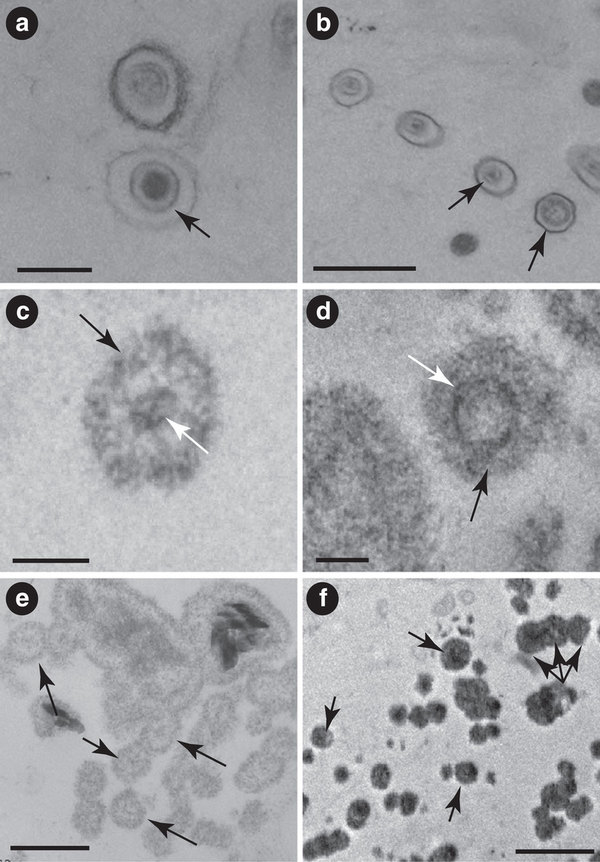
The bacteriophage they used is the Salmonella phage P22.  P22’s capsid is an icosahedron composed of a major coat protein assembled onto a scaffold protein. Coat protein and scaffold protein self-assemble into a procapsid that during a normal infection is then packaged with the phage DNA.  The researchers fused the two subunits of a hydrogenase protein to separate scaffold protein genes.  When expressed together with coat protein, procapsid self-assembled with the heterodimeric hydrogenase protein inside as outlined in this figure.

Hyman blog post

(modified from Jordan 2015)

When they tested for hydrogenase activity, the highest efficiency was found if the scaffold protein/hydrogenase subunits were expressed several hours before the coat protein (line one in the table).  This pre-encapsulation period presumably allowed the two hydrogenase subunits to assemble into the active heterodimer before being constrained by the coat protein.

pH 5 pH 8
P22 encapsulated with sequential expression 6118 nmol H2/mg min 3218 nmol H2/mg min
P22 encapsulated with simultaneous expression 757 nmol H2/mg min
Unencapsulated hydrogenase + scaffold without coat protein 46 nmol H2/mg min 12.6 nmol H2/mg min
Free hydrogenase 12-38 nmol H2/mg min (pH not specified)

Data from Jordan 2015

Additional experiments showed that the encapsulation also partially protected the hydrogenase against trypsin, heat denaturation (60°C for 45 min.) and air exposure.

The reason for the increased enzymatic activity is not entirely clear.  The increased activity and protection results suggest that the enzyme’s quaternary structure is stabilized in some way in the capsid.  It may also be that enzyme efficiency is higher when several hundred copies of the enzyme are in close proximity to each other in some sort of synergistic effect.

Overall, this paper demonstrates another way that phages can be used in non-biological technologies as well as biological.  Independent of the phage aspect, an improved catalyst for production of hydrogen gas could prove quite valuable as alternative fuels, such as hydrogen, are increasingly sought after.

Reference: Paul C. Jordan, Dustin P. Patterson, Kendall N. Saboda, Ethan J. Edwards, Heini M. Miettinen, Gautam Basu, Megan C. Thielges, and Trevor Douglas, “Self-Assembling Biomolecular Catalysts for Hydrogen Production”, Nature Chemistry doi:10.1038/nchem.2416, published on-line December 21, 2015.

The ecology of viruses that infect eukaryotic algae

The world of algal viruses has only gotten more interesting in recent years as it has become increasingly clear just the sheer quantity of mortality they cause and how much that drives global nutrient cycling in ways we’ve been largely blind to.

Chlorovirus

The ecology of viruses that infect eukaryotic algae

Because viruses of eukaryotic algae are incredibly diverse, sweeping generalizations about their ecology are rare. These obligate parasites infect a range of algae and their diversity can be illustrated by considering that isolates range from small particles with ssRNA genomes to much larger particles with 560 kb dsDNA genomes. Molecular research has also provided clues about the extent of their diversity especially considering that genetic signatures of algal viruses in the environment rarely match cultivated viruses. One general concept in algal virus ecology that has emerged is that algal viruses are very host specific and most infect only certain strains of their hosts; with the exception of viruses of brown algae, evidence for interspecies infectivity is lacking. Although some host–virus systems behave with boom-bust oscillations, complex patterns of intraspecies infectivity can lead to host–virus coexistence obfuscating the role of viruses in host population dynamics. Within the framework of population dynamics, host density dependence is an important phenomenon that influences virus abundances in nature. Variable burst sizes of different viruses also influence their abundances and permit speculations about different life strategies, but as exceptions are common in algal virus ecology, life strategy generalizations may not be broadly applicable. Gaps in knowledge of virus seasonality and persistence are beginning to close and investigations of environmental reservoirs and virus resilience may answer questions about virus inter-annual recurrences. Studies of algal mortality have shown that viruses are often important agents of mortality reinforcing notions about their ecological relevance, while observations of the surprising ways viruses interact with their hosts highlight the immaturity of our understanding. Considering that just two decades ago algal viruses were hardly acknowledged, recent progress affords the optimistic perspective that future studies will provide keys to unlocking our understanding of algal virus ecology specifically, and aquatic ecosystems generally.

Data Storage and Standard Parts

Genetic data storage, scaleable cell-cell communication, and still-better gene expression, all thanks to phage!

Cross-talk between Diverse Serine Integrases

Abstract
Phage-encoded serine integrases are large serine recombinases that mediate integrative and excisive site-specific recombination of temperate phage genomes. They are well suited for use in heterologous systems and for synthetic genetic circuits as the attP and attB attachment sites are small (< 50 bp), there are no host factor or DNA supercoiling requirements, and they are strongly directional, doing only excisive recombination in the presence of a recombination directionality factor. Combining different recombinases that function independently and without cross-talk to construct complex synthetic circuits is desirable, and several different serine integrases are available. However, we show here that these functions are not reliably predictable, and we describe a pair of serine integrases encoded by mycobacteriophages Bxz2 and Peaches with unusual and unpredictable specificities. The integrases share only 59% amino acid sequence identity and the attP sites have fewer than 50% shared bases, but they use the same attB site and there is non-reciprocal cross-talk between the two systems. The DNA binding specificities do not result from differences in specific DNA contacts but from the constraints imposed by the configuration of the component half-sites within each of the attachment site DNAs.

New Applications for Phage Integrases

Within the last 25 years, bacteriophage integrases have rapidly risen to prominence as genetic tools for a wide range of applications from basic cloning to genome engineering. Serine integrases such as that from ϕC31 and its relatives have found an especially wide range of applications within diverse micro-organisms right through to multi-cellular eukaryotes. Here, we review the mechanisms of the two major families of integrases, the tyrosine and serine integrases, and the advantages and disadvantages of each type as they are applied in genome engineering and synthetic biology. In particular, we focus on the new areas of metabolic pathway construction and optimization, biocomputing, heterologous expression and multiplexed assembly techniques. Integrases are versatile and efficient tools that can be used in conjunction with the various extant molecular biology tools to streamline the synthetic biology production line.

21st Biennial Evergreen International Phage Meeting!

the-evergreen-state-college

You can register for the 21st Biennial Evergreen International Phage Meeting Aug. 2-7 on the Evergreen web site: www.evergreen.edu/phage.  If you register by April 30, you will qualify for the “Early Registration” rate: $600 for academics, $700 for Corporate Rate, $450 for graduate and undergraduate students and guests.  This covers all meeting costs, including room and board.  (If you choose to stay off campus, it will be $150 less, but still include meals.)  Some registration assistance is potentially available.

You will then have until May 24 to at least pay a $100 nonrefundable deposit, apply for assistance, or tell us that you are waiting for a visa.  If you register after May 1, the rates will be $100 more in each category.  Further meeting information is on the web site and will be regularly updated there.

Another layer of understanding to add to the genome injection story

Releasing the packaged viral DNA into the host cell is an essential process to initiate viral infection. In many double-stranded DNA bacterial viruses and herpesviruses, the tightly packaged genome is hexagonally ordered and stressed in the protein shell, called the capsid. DNA condensed in this state inside viral capsids has been shown to be trapped in a glassy state, with restricted molecular motion in vitro. This limited intracapsid DNA mobility is caused by the sliding friction between closely packaged DNA strands, as a result of the repulsive interactions between the negative charges on the DNA helices. It had been unclear how this rigid crystalline structure of the viral genome rapidly ejects from the capsid, reaching rates of 60,000 bp/s. Through a combination of single-molecule and bulk techniques, we determined how the structure and energy of the encapsidated DNA in phage λ regulates the mobility required for its ejection. Our data show that packaged λ-DNA undergoes a solid-to-fluid–like disordering transition as a function of temperature, resulting locally in less densely packed DNA, reducing DNA–DNA repulsions. This process leads to a significant increase in genome mobility or fluidity, which facilitates genome release at temperatures close to that of viral infection (37 °C), suggesting a remarkable physical adaptation of bacterial viruses to the environment ofEscherichia coli cells in a human host.

Significance

The efficiency of viral replication is limited by the ability of the virus to eject its genome into a cell. We discovered a fundamentally important mechanism for translocation of viral genomes into cells. For the first time, to our knowledge, we show that tightly packaged DNA in the viral capsid of a bacterial virus (phage λ) undergoes a solid-to-fluid–like structural transition that facilitates infection close to 37 °C. Our finding shows a remarkable physical adaptation of bacterial viruses to the environment of Escherichia coli cells in a human host.

In the beginning…

Young_d'Herelle“From a modern microbiological viewpoint the motivation behind the experiments that led to the discovery of bacteriophage is hard to understand. What was the purpose of filtering a bacterial culture to remove the bacteria, then remixing the filtrate with a fresh bacterial culture? We all know the outcome of this classic experiment, but why was it done in the first place? To fathom the intent of the original investigator, Félix d’Herelle, one has to put aside twenty-first century ideas and recover the context of microbiology and disease in the early twentieth century.

An M13 phage based colorometric sensor

Having previously demonstrated the ability to build collagen-like self-templating supramolecular structures out of M13 phage filaments, this year the Seung-Wuk Lee Lab has come out with a new paper demonstrating their ability to use those structures as a litmus to sensitively detect things like humidity, volatile organic compounds, or TNT explosives and report color strongly enough for an iPhone camera to distinguish:

Biomimetic virus-based colourimetric sensors
Many materials in nature change colours in response to stimuli, making them attractive for use as sensor platform. However, both natural materials and their synthetic analogues lack selectivity towards specific chemicals, and introducing such selectivity remains a challenge. Here we report the self-assembly of genetically engineered viruses (M13 phage) into target-specific, colourimetric biosensors. The sensors are composed of phage-bundle nanostructures and exhibit viewing-angle independent colour, similar to collagen structures in turkey skin. On exposure to various volatile organic chemicals, the structures rapidly swell and undergo distinct colour changes. Furthermore, sensors composed of phage displaying trinitrotoluene (TNT)-binding peptide motifs identified from a phage display selectively distinguish TNT down to 300 p.p.b. over similarly structured chemicals. Our tunable, colourimetric sensors can be useful for the detection of a variety of harmful toxicants and pathogens to protect human health and national security.

rsz_1ncomms4043-f1
“Bioinspired phage-based colourimetric sensors, termed Phage litmus, are composed of hierarchical bundles like the collagen fibres in turkey skins. Application of target molecules (chemical stimuli) causes colour shifts due to structural changes, such as bundle spacing (d1 and d2) and coherent scattering. Using a handheld device’s camera (iPhone) and home-built software (iColour Analyser), we can identify target molecules in a selective and sensitive manner.”

Fossilized viruses found in the geological record

In the 90s there was a notion that was inexplicably popular in the literature (Folk, 1993; Folk and Lynch, 1997; Pedone and Folk, 1996; Sillitoe et al., 1996; McKay et al., 1996; Vasconcelos and McKenzie, 1997; Kajander and Çiftçioglu, 1998), and still stubbornly held by some, that the distinct structured particles under 150 nm in size commonly found in the geological record associated with putatively biogenic rock constituted a new form of “nanobacteria,” despite being too small to support living systems and based on often poorly labelled micrographs. How much the controversy this engendered unknowingly recalled the classical extended fights over Martinus Beijerinck‘s contagium vivum fluidum and the original discovery of viruses may seem obvious only in hindsight, but a new paper now convincingly argues that many of these particles may indeed be fossilized viruses of microbes,

Viruses as new agents of organomineralization in the geological record

Muriel Pacton, David Wacey, Cinzia Corinaldesi, et al. Published 2014 in Nat. Commun. doi:10.1038/ncomms5298
Viruses are the most abundant biological entities throughout marine and terrestrial ecosystems, but little is known about virus–mineral interactions or the potential for virus preservation in the geological record. Here we use contextual metagenomic data and microscopic analyses to show that viruses occur in high diversity within a modern lacustrine microbial mat, and vastly outnumber prokaryotes and other components of the microbial mat. Experimental data reveal that mineral precipitation takes place directly on free viruses and, as a result of viral infections, on cell debris resulting from cell lysis. Viruses are initially permineralized by amorphous magnesium silicates, which then alter to magnesium carbonate nanospheres of ~80–200 nm in diameter during diagenesis. Our findings open up the possibility to investigate the evolution and geological history of viruses and their role in organomineralization, as well as providing an alternative explanation for enigmatic carbonate nanospheres previously observed in the geological record.

While the authors seem primarily concerned with how viruses could affect the chemical formation of geological features, if real, this has the exciting potential to allow us to track the evolution of viral morphology through geological time.

Figure 2(a) Virus-like particle characterized by an icosahedral capsid-like structure (black arrow). Scale bar, 200 nm. (b) Virus-like particles. Black arrows point to the capsid-like structure in each case. Scale bar, 500 nm. (c) First stage of the amMg-Si mineralization process of the icosahedral capsid-like structure (black arrow); white arrow points to the viral DNA inside. Scale bar, 100 nm. (d) Second stage of the amMg-Si mineralization process of a virus-like particle (black arrow) showing its icosahedral capsid-like structure (white arrow). Scale bar, 100 nm. (e) Early mineralization of virus-like particles showing amMg-Si permineralized capsid-like structures (arrows). Scale bar, 200 nm. (f) amMg-Si permineralized virus-like particles occurring as single entities and chains (examples arrowed). Scale bar, 500 nm.